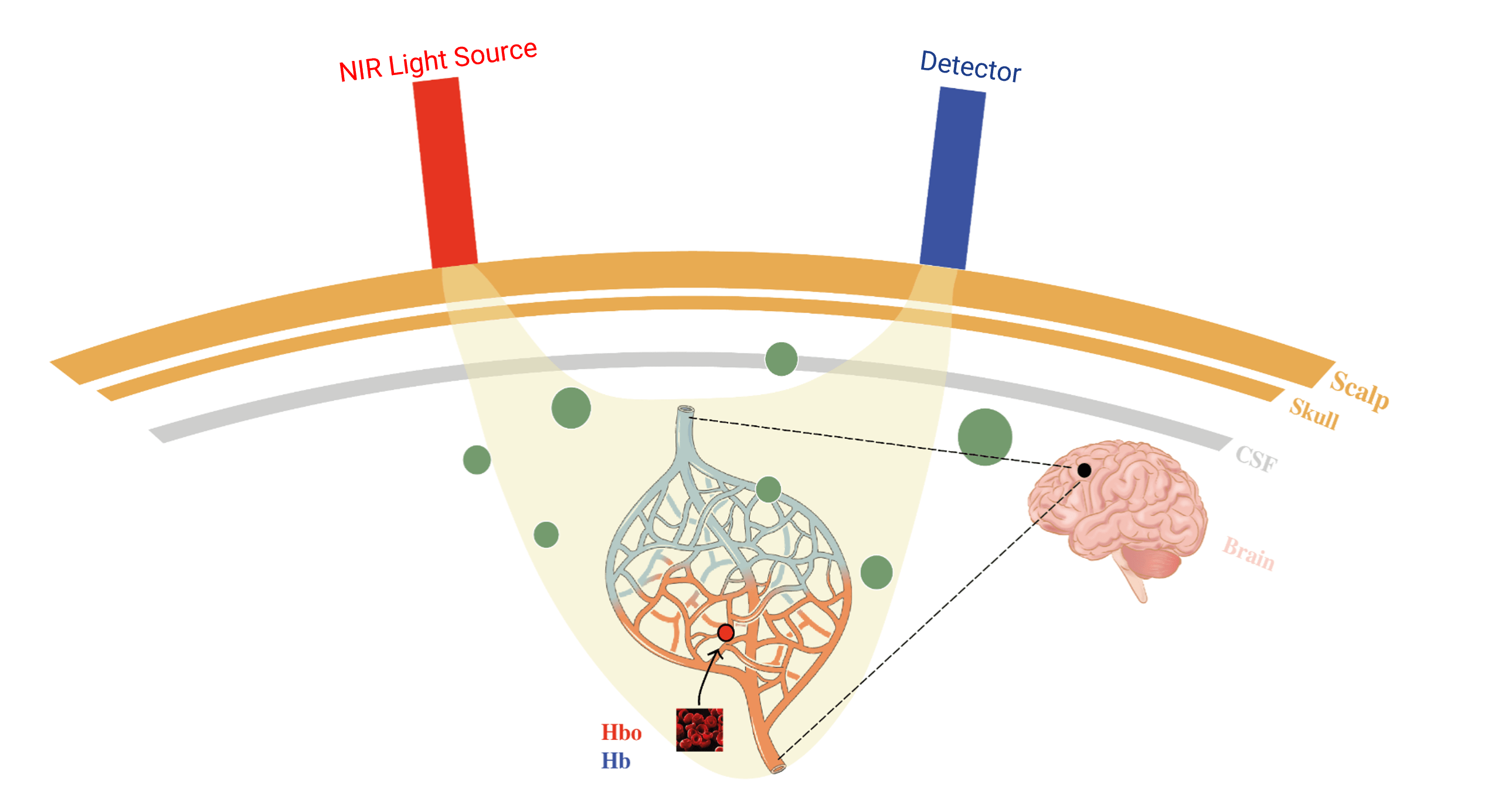
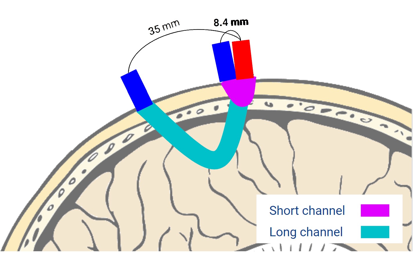
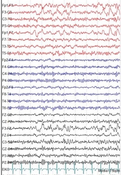
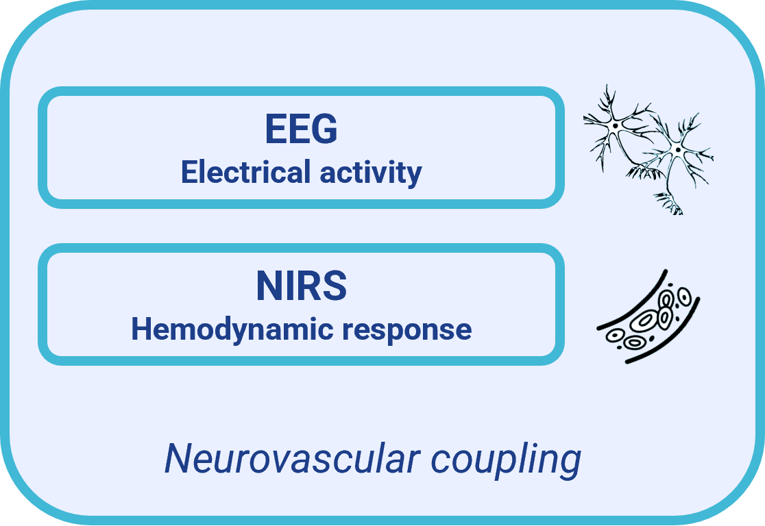
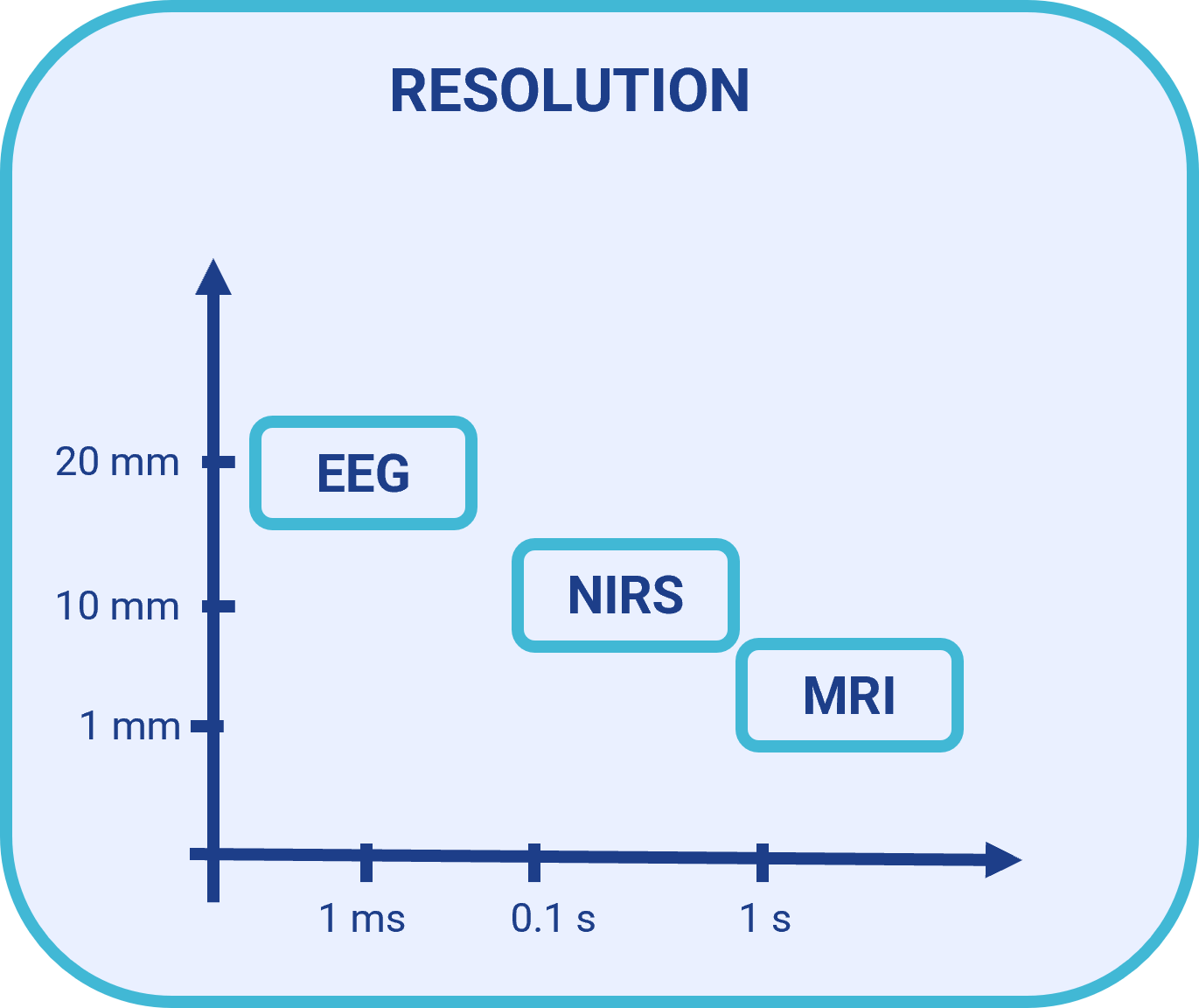
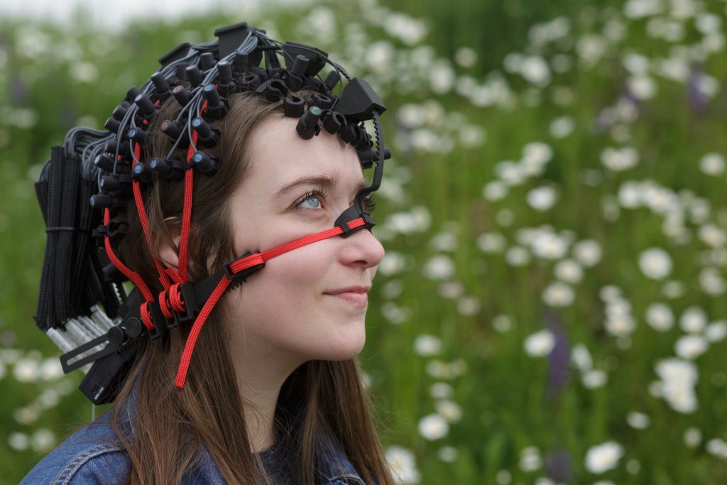
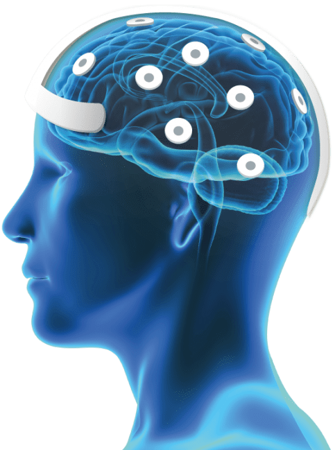
SEENEL IMAGING
Research-based from INSERM lab, Seenel Imaging commercializes Medelopt®, a breakthrough neuroimaging device.
Developed by world-renowned clinical neuroscience and signal processing researchers, along with a well-rounded team of electronic onboarding
and medical devices experts, Medelopt® is the perfect multimodal functional brain exploration platform for neuroscientific researchers.
OUR TECHNOLOGY
1. Schematic diagram of the functional NIRS (fNIRS). NIR-lights are generated and guided to the human’s head via optical fibers or cables. Another fiber bundle or cable directs diffusively reflected light from the head to detectors. A light detector captures the light resulting from the interaction with the chromophores (e.g. HbO, HbR), following a crescent-shaped path back to the surface of the skin. fNIRS takes advantage of the living tissue’s absorbing properties in the near-infrared range to measure changes in the local concentrations of oxy- and deoxy-hemoglobin ([HbO] and [HbR]) through intact skulls. Light in the NIR spectral range (650–950 nm ‘optical window’) can penetrate relatively deeply (a few centimeters) into the brain tissue, mostly because NIR light is only slightly absorbed by water, collagen, and proteins. fNIRS is performed by emitting near-infrared light into the scalp and detecting the transmitted light at certain positions.
2. EEG measures the sum total activity of the electrical signals that the brain sends to the scalp. EEG signals – as distinct from the fNIRS, which measures brain activity indirectly (i.e., changes in oxygenation) – are therefore generated directly through neuronal activity. The fNIRS’s spatial resolution is better than the EEG’s, and the EEG’s temporal resolution is better than the fNIRS’s

Research-based**
(INSERM U1105/UPJV/CHU Amiens Picardy)
and protected by 3 patents,
Medelopt® is a breakthrough innovative multimodal
functional cerebral neuroimaging device.
Medelopt® provides high quality measurements
of changes in oxy and deoxy-haemoglobin
simultaneously with electric potentials.
**Safaie.J et al. (2013)
KEY FEATURES

High density bimodality
fNIRS/EEG
Hemodynamic (with 512 channels) & featuring electrical signal acquisition, Medelopt® provides accurate measurements for in-vivo 2D/3D functional brain mapping.
LED: 16 emitters / Electropods: 32 receptors / 8 EEG electrodes

Sophisticated technology
crafted for Wearability
Medelopt® miniaturisation allows acquisitions on moving patients.

Designed for getting direct access
to the scalp
Due to its unique design structure, Medelopt® solves the hair constraint which is the main fNIRS-related constraint that researchers encounter. LED-emitters LED and electropods come directly in contact with the scalp’s skin.

Customized
headset setup
Thanks to the headset design, sensor placement (emitters/Receptors and EEG electrodes) can be set up to record the whole head, from the cognitive to the visual area.

Long recording
is now possible
Pressure zones are distributed homogeneously and uniformly around the head. Because of this, long recordings are made possible.
APPLICATIONS

SPORT SCIENCES
The possibility given by NIRS to move freely makes possible recordings of
brain activity during outdoor activities or physical exercise.
Exploring brain mechanisms involved in sport sciences
improve our understanding of the relationship
between mind and motor action or the effect of cognition on motor performance
during localized exercise or whole-body exercise. For example, NIRS devices could
be a way to explore decision-making mechanisms in reactionary sport like
tennis or to determine how physical exercise influence the main executive functions
COGNITIVE NEUROSCIENCE
The point of a wearable device for cognitive neuroscience that evaluates brain function in response to sensory stimulation lies essentially in the possibility of developing ecological stimuli that minimic everyday life situations in the best way possible. With a wearable device, it becomes possible to map out the brain structures involved in tasks such as driving or sports in real-life situations, as opposed to what is possible with fMRIs.
DBS/VSN
The appeal of the device lies in its compatibility with devices that involve electrical stimulation of the central (Deep Brain Stimulation (DBS)) or peripheral (Vagus Nerve Stimulation (VSN)) nervous system. This device thus makes it possible to map out the structures of the cortical surface that are modulated by these stimuli without having them be generated within the constraints of the electric fields produced by the stimulation or the constraints of magnetic fields inherent to MRIs and that make these maps difficult to produce in cases of DBS and VNS.

Video of the dynamic appearance of NIRS results in a patient with epilepsy with a central initial focus that spreads in the frontal regions during an epileptic interictal spike.
EPILEPSY RESEARCH
Epilepsy involves complex mechanisms that combine neuronal, astrocytic, and vascular interactions, particularly within the neurovascular coupling involved in epileptic spikes and seizures.
The simultaneous analysis of various neuronal and vascular compartments by the EEG in tandem with the NIRS makes it possible to see the mechanisms involved and their interactions by a multimodal, multidimensional approach. Additionally, this simultaneous approach combines the EEG’s high temporal resolution with the high spatial resolution of the NIRS
WHO WE ARE
CONTACT
CUSTOMERS & PARTNERSHIPS
We are proud to provide world-leading functional neuro-imaging research product and to build partnership with
.
.
.
.